Both a general interest in Optics and Electro Optics as well as an interest in spy related technologies like cryptography (Spy Nickel) aroused my interest in simple microscopes (Wiki). A Compound microscope (Wiki) is the type most people know and uses both an objective lens and an eyepiece whereas the simple microscope has no eyepiece. It's similar to a magnifying glass (Wiki) or loupe (Wiki) except with much higher power.
Examples of compound microscopes are: Nikon Labophot, Nikon SMZ-U Stereo, Mitutoyo Toolmakers Measuring Microscope, Unitron No. 83444 Microscope, Unitron Auto-Illumination Inverted Microscope, Bausch & Lomb StereoZoom. All of these make use of separate objective and eyepiece lens assemblies.
This is a project at Stanford (Prakash Labs) to make a very low cost microscope that can be used in developing countries for medical testing. Since there are a number of tests that now require a lab quality microscope and someone trained in it's use those tests take a long time because the microscopes are very expensive and there are not very many of them. A very low cost microscope will enable more people to learn how to do the tests and the testing can be done locally. There might be different versions of the Foldscope for different tests or some combination of different components like is done on lab microscopes.
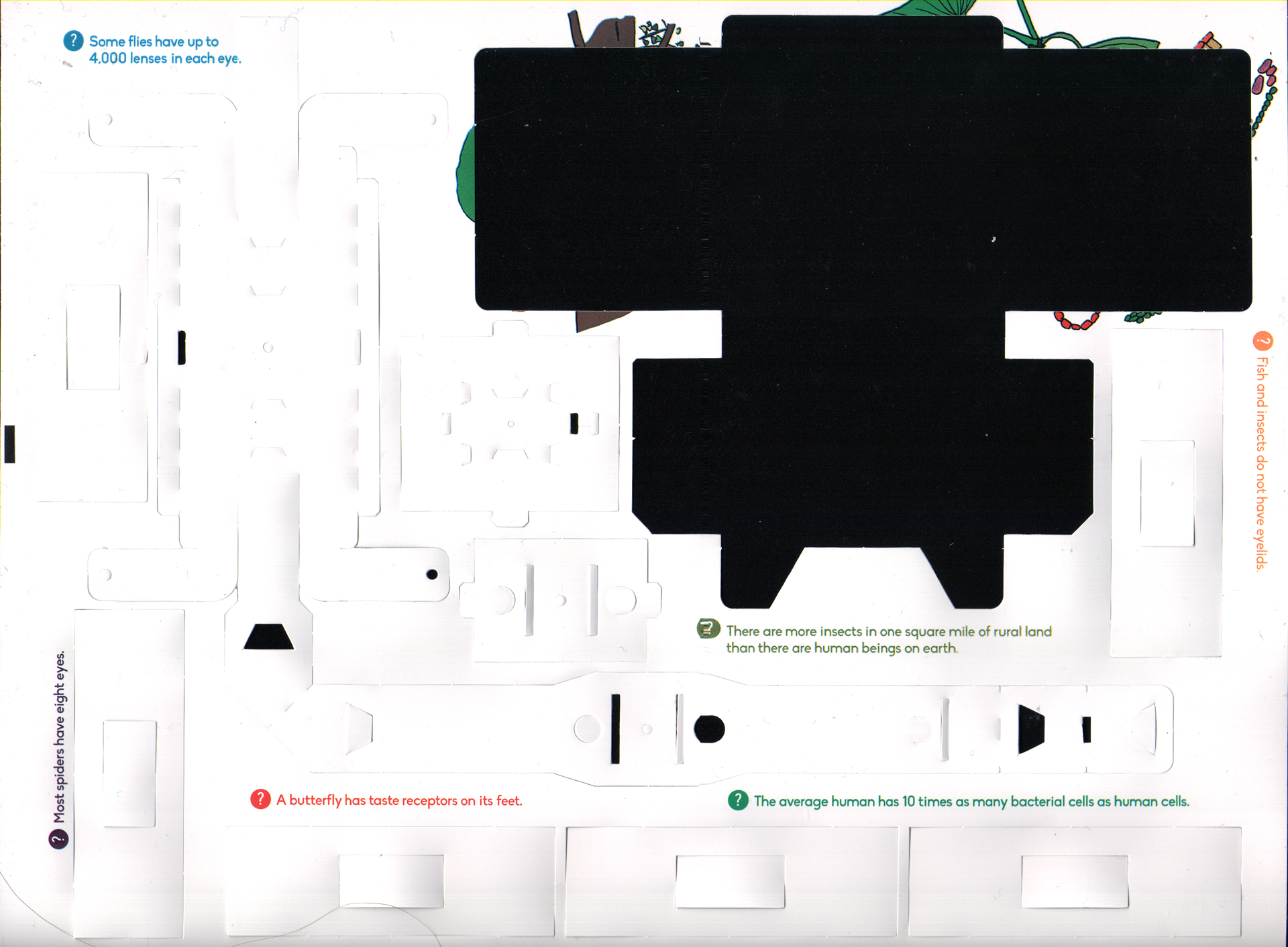
The Foldscopes I just received (Dec. 2014) consist of two sheets of tag board which are identical, but there are different lenses and one optional LED illuminator. So the basic Foldscope can be configured with two different objectives and either no built-in illumination or a white LED illumination. Maybe there will be a future version with a UV light source and filters for fluorescence microscopy.
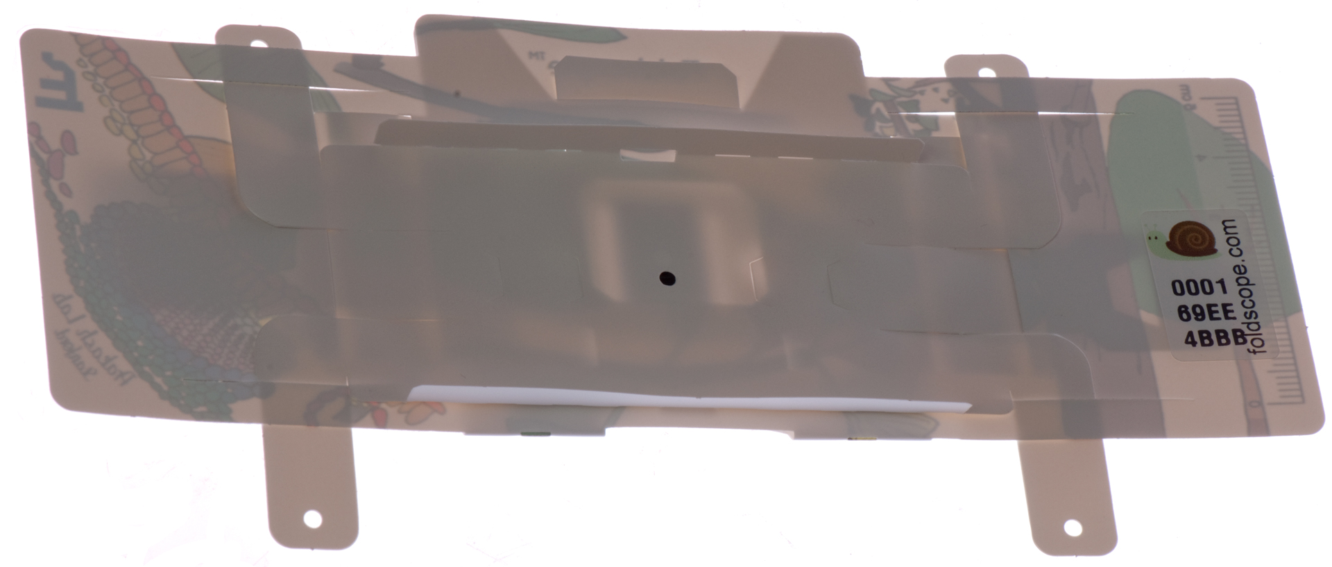
The Foldscopes arrived in a standard 12-1/2" x 9-1/2" envelope that weighs less than 5 ounces. This is a very important consideration since shipping adds to the cost of the product. If shipped in bulk you can subtract the envelope weight of 1.7 oz. resulting in a net weight of close to 3 oz for this kit and more like a little over 1 oz per Foldscope.
The paper parts that come on the 8-1/2" x 11" sheet are really more like a combination of plastic and paper. They appear to be waterproof.
Reference Designations of parts
See Fig 2 & Fig 3
Designation
Description
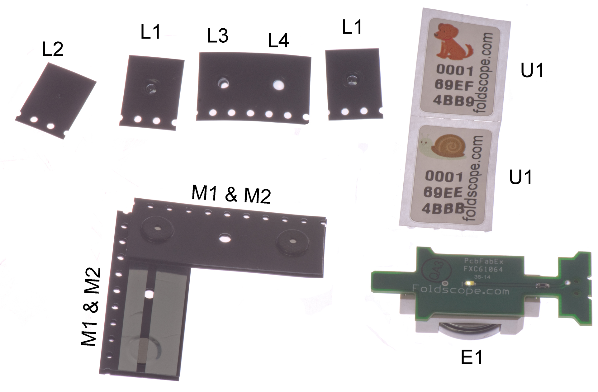
E1
Light Module (LED & CR2032 battery, On/Off switch)
L1
Low Magnification Lens
small aperture, large lens
L2
High Magnification Lens
Very small aperture, small lens
L3
High Mag Lens holder
large aperture, no lens
L4
Condenser Lens
large aperture, large lens
M1
Magnet strip for FoldScope
black adhesive tape
M2
Magnet strip for phone
silver adhesive tape
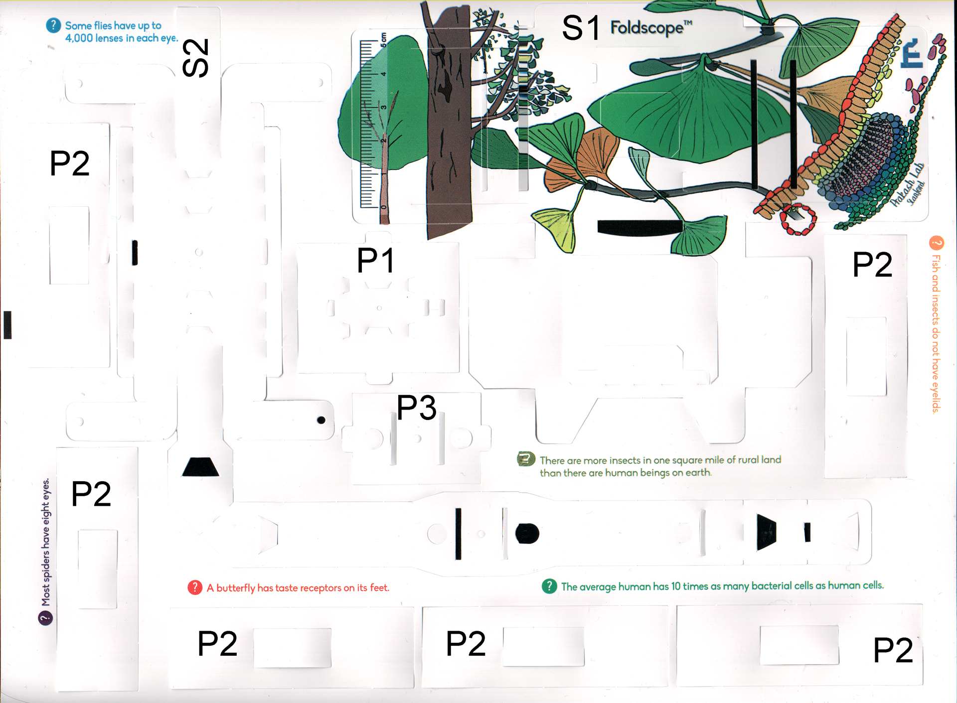
P1
Paper LED Light Module holder
P2
Paper slide (6 ea/Foldscope)
P3
Paper Magnet holder
S1
Main Stage
S2
Sliding Stage
T1
Transparent Tape Stickers (sheet of 60/FoldScope)
T2
0.2" wide double sided Tape (8"/FoldScope)
U1 & U2
Unique serial number Label for each Foldscope
Photos
Note movie film has been around for a long time and was adopted by the electronics Surface Mount business to hold parts. The foldscope uses movie film as a packaging method for the ball lenses.
Fig 1 Mailing Envelope
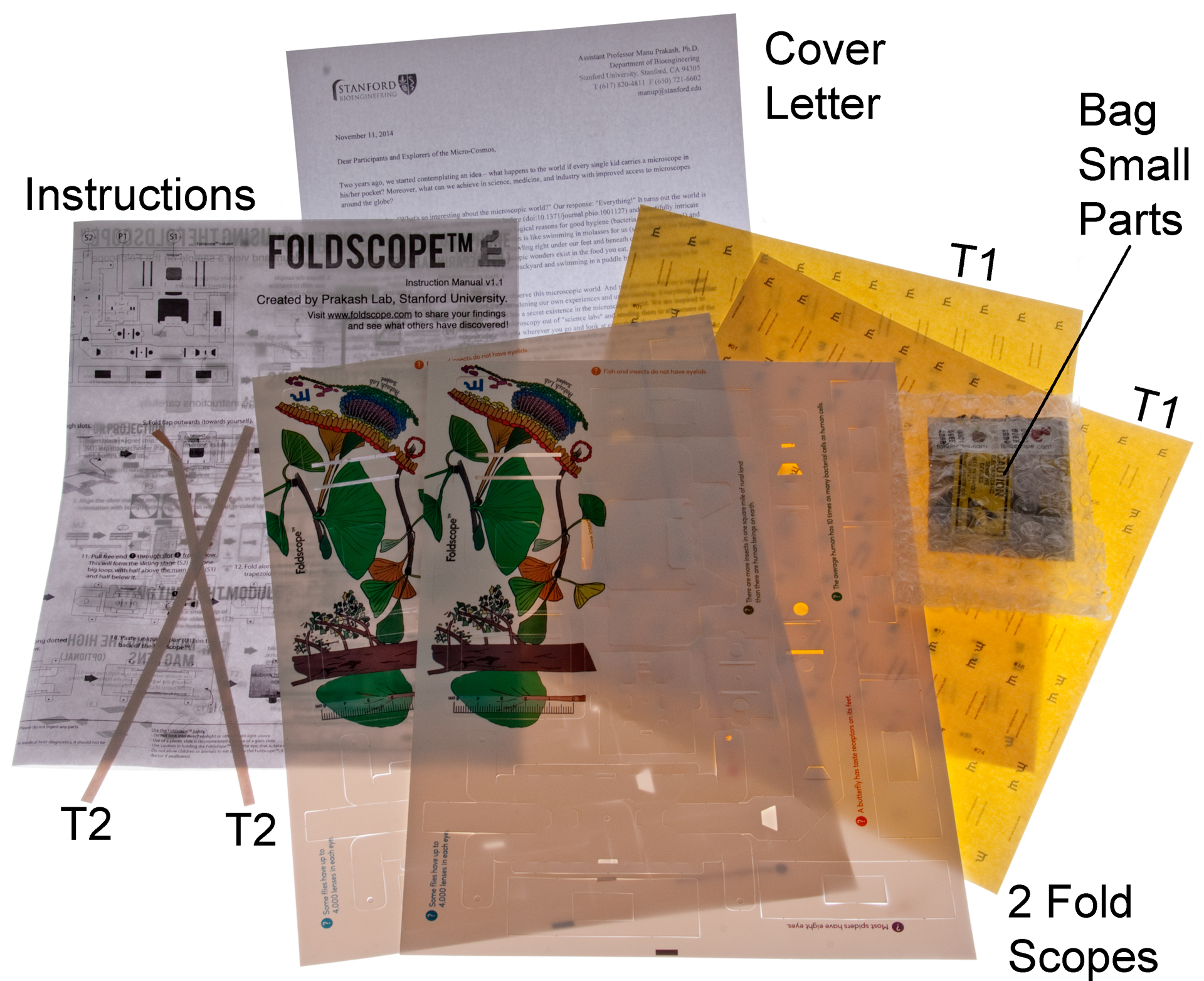
Fig 2 In the envelope
Fig 3 In the Bag of Parts
Fig 4 a FoldScope before separating the S & P parts
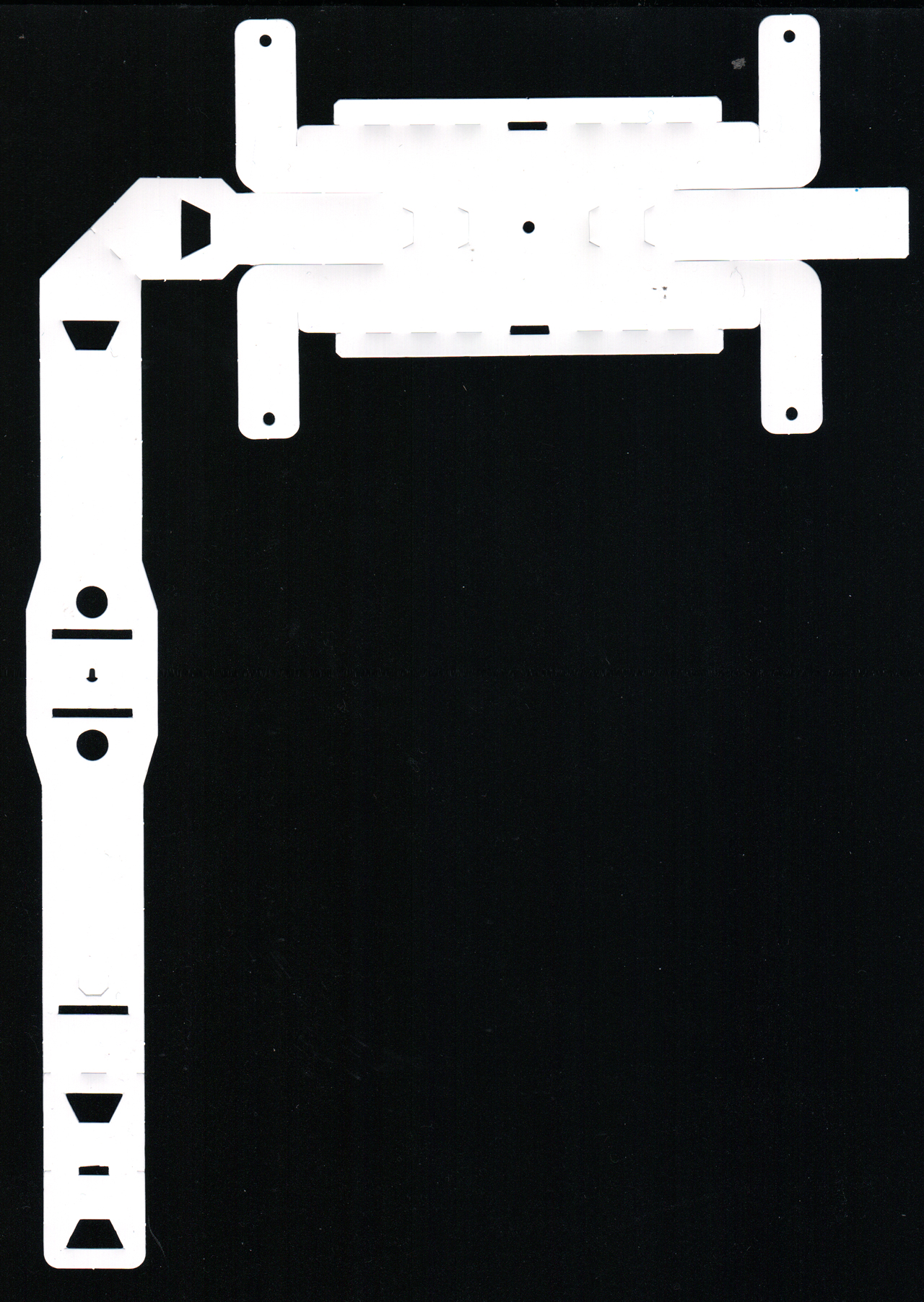
Fig 5 after removing S1 Main Stage
Fig 6 S1 Main Stage flat
Fig 7 S2 Sliding Stage flat
Fig 8 Eyepiece side
Note: at right the S2 tab has come out of the top part of S2 so the condenser hole on the back is not tracking the objective on the front. Easy to fix, just put it in the slot on the back of the front part.
Fig 9 Illumination side
Operation
The main stage (S1) is held in the left and right hands using the middle and index fingers. The sliding stage (S2) is held between the thumbs and index fingers. Relative motion between the main stage (S1) and sliding stage (S2) allows you to scan the slide to locate the subject.
Note: All 4 of the the vertical tabs of the sliding stage should move when the sliding stage is moved. If the right hand tab on the square end of the sliding stage becomes disconnected then the light hole on the back side will not track the objective on the front side.
In order to see there needs to be a direct source of light like looking directly at a desk lamp. Looking at a white wall does not work.
When the LED Light module and condenser lens are used the light is very bright and fine detail can be seen.
To focus you can pull apart on the S1 main stage or pull apart on the S2 sliding stage and/or bend the main or sliding stage. You can focus on different parts of an insect leg and see a lot of detail, but not with as wide a field of view as on a compound microscope like the Nikon Labophot.
Options
High-Mag Lens
Uses the L2 lens inside the L3 holder.
Smart Phone magnetic holder
Used with the M1 and M2 magnetic attachments on front
Smart Phone projection illumination
Used with the M1 and M2 magnetic attachments on back
LED Light Module
Used with the P1 Paper LED holder and the L4 condenser Lens.
Technical Details
This is by far the best technical description including details on not only this bright field version, but also on other versions.
POLS - Foldscope: Origami-Based Paper Microscope - Full document.pdf - Supplementary data.docx
Strehl ratio (Wiki) is part of the second resolution metric (RM2) in the supplement.
A Better Method of Measuring Optical Performance: Move over P-V and make way for Strehl
Third-order aberration coefficients of a thick lens Antonin Mikš and Jiří Novák, Applied Optics, Vol. 51, Issue 33, pp. 7883-7886 (2012)
Also see Ball Lens below.
A new 3D printed microscope from Stanford that includes a PacMan type game on an Android smart phone. Can be done using a conventional compound microscope or using a 3D printed microscope. “Ludus,” which means “play,” “game” or “elementary school.”
Stanford: Smartphone microscope developed by Stanford bioengineer creates interactive tool for microbiology - pdf - Supporting Info -
"The LudusScope can be adapted to fit onto a standard microscope. (B) A 3D printable microscope attachment is needed (S2 Note). The same 3D printed sample holder as the full version can be used, as well as the same circuit sans illumination LED. This alternative approach may be more convenient for classrooms that already have access to standard microscopes.
PLOS Journal: LudusScope: Accessible Interactive Smartphone Microscopy for Life-Science Education -
Antonie van Leeuwenhoek (Wiki) was born in the same week as Johannes Vermeer (Wiki) and they spent most of their lives living on the town square of Delft. I discovered this while learning about the Hockney–Falco thesis (Wiki, Optics-Art.com). It seems as a settled fact that Vermeer used optics to get photo realistic painting and there were other "Dutch Masters" that did the same. The book Eye of the Beholder (Ref 4) goes into a lot of detail about the lives of both Leeuwenhoek and Vermeer. For many years Leeuwenhoek would send observation reports to the Royal society but would not divulge his methods. This was in the 1600s when "Guilds" were common and kept secret the methods used by artisans. That is part of an explanation why Vermeer did not disclose how he made photo realistic paintings. See the movie: Tim's Vermeer to see a method that probably captures the major points of Vermeer's method.
Found this on the Quekett web page Replica Leeuwenhoek Microscopes which includes an email address for Chris Allen (gillandchris@btinternet.com).
Google - Antoni van Leeuwenhoek’s 384th Birthday - October 24, 2016
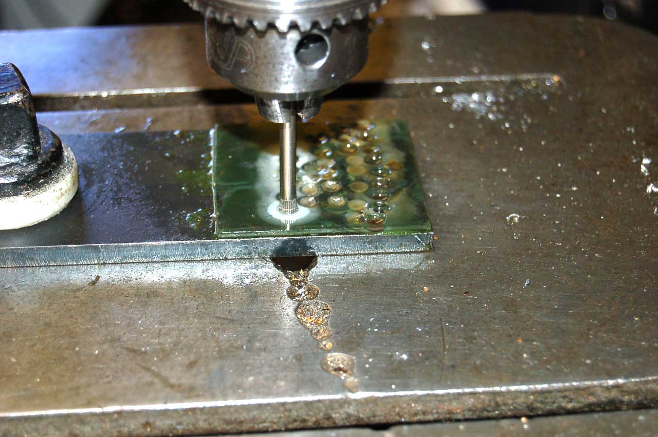
He makes the lens by drilling a cylinder of common window glass, manually grinding it into rough shape, then finish grinding to get the desired spherical radius on each side then polishing.
YouTube:Through van Leeuwenhoek’s Eyes: Microbiology in a Nutshell - it's not clear how she looked at liquid s. Dr. Lesley A. Robertson at Delft. At the beginning there's a rotating simple microscope with what appears to be a disk specimen holder. This may be a pair of mica disks with pond water between them?
Google Books: Antoni van Leeuwenhoek: Master of the Minuscule - some is visible on Google Books. Over $100 from Amazon.
R1 = R2 = 2.55mm. About 1.7mm thick with a refractive index around 1.5.
Plugging those numbers into the Lens Maker's equation (Wiki) gives:
1/f = (1.5-1.0) [1/2.55 + 1/2.55 + ((1.5 - 1.0)*1.7)/(1.5*2.55*2.55) = 0.5 *0.87146 = 0.43573
f= 2.295mm or 436 diopters (Wiki)
Power = 0.25m * diopters +1 = 110 power. (Note: 0.25 meters (10") is about the normal distance from a book to your eye, i.e. normal eye focal distance.)
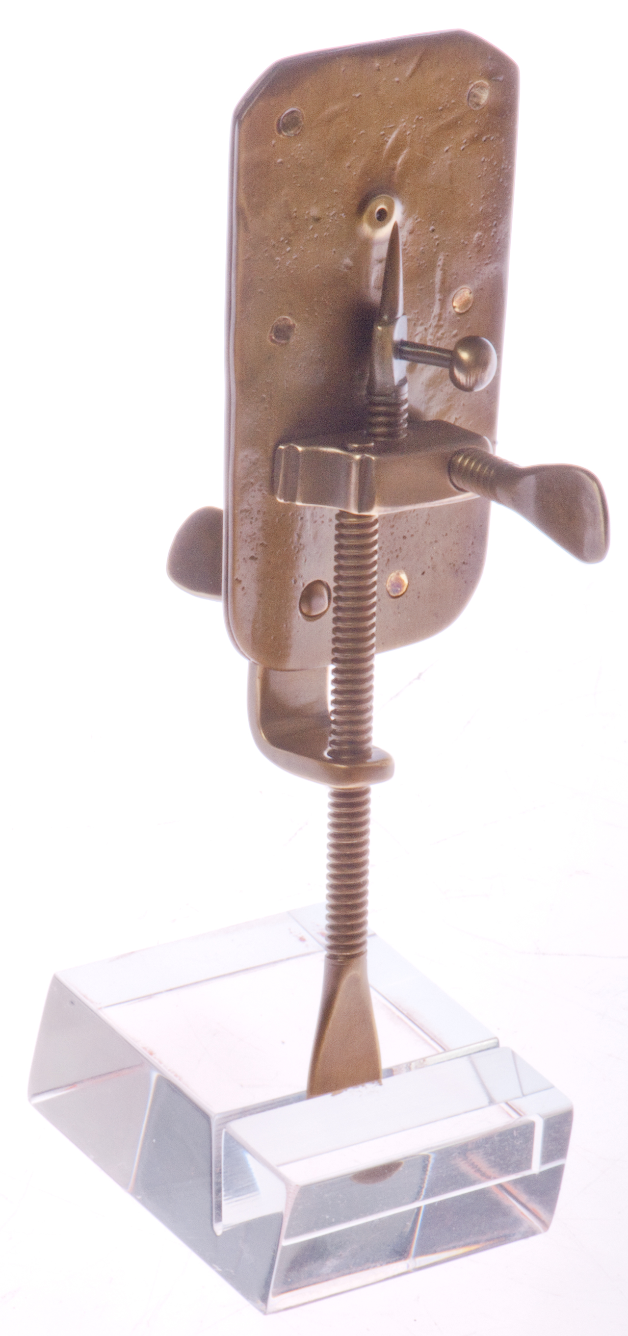
Using a Simple Microscope
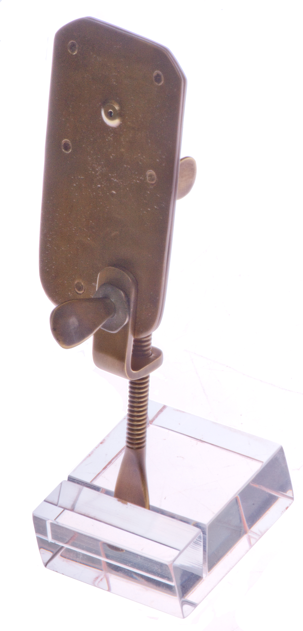
Note when these were in use they didn't have slides so instead you put the thing you wanted to see on the point.Note 1
The ball ended adjustment lets you rotate the object being viewed and raises or lowers it a little.
The short screw on the subject side is to change the focus distance.
The long screw that's supporting the microscope on the stand is for moving the subject up or down.
The pinch screw on the front allows tilting the subject left or right of the lens.
It would make a nice addition to have a way to remove the pinch screw and replace the old mechanism with one that would accept a standard 1" x 3" glass microscope slide.
Note 1: In the book Eye of the Beholder (Ref 4) there are a number of references to Leeuwenhoek using glass capillary tubes (Wiki) to hold the liquid samples he was observing. A glass tube can be heated then pulled to make extremely fine tubing (Wiki). I wonder if such a tube would act as a lens to view the water inside it?
The "eel glass" looks like it holds a standard test tube which held an eel. But if used for pond water would allow way too much front to back motion so not able to focus.
Photos
Fig 1 Eye side
Fig 2 Subject side
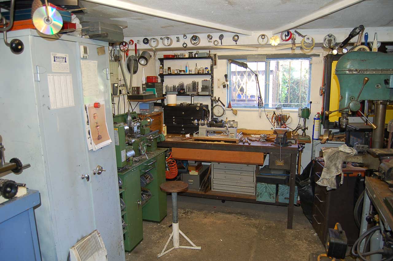
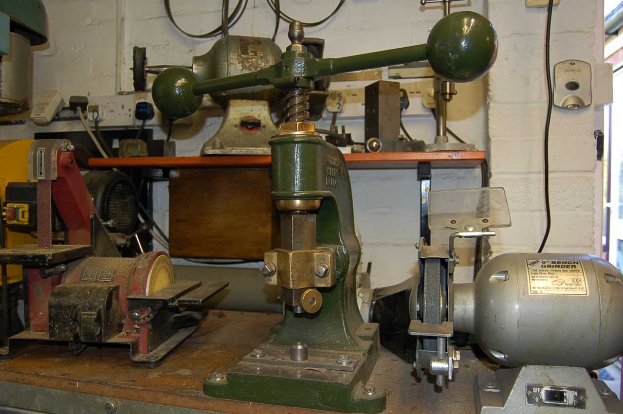
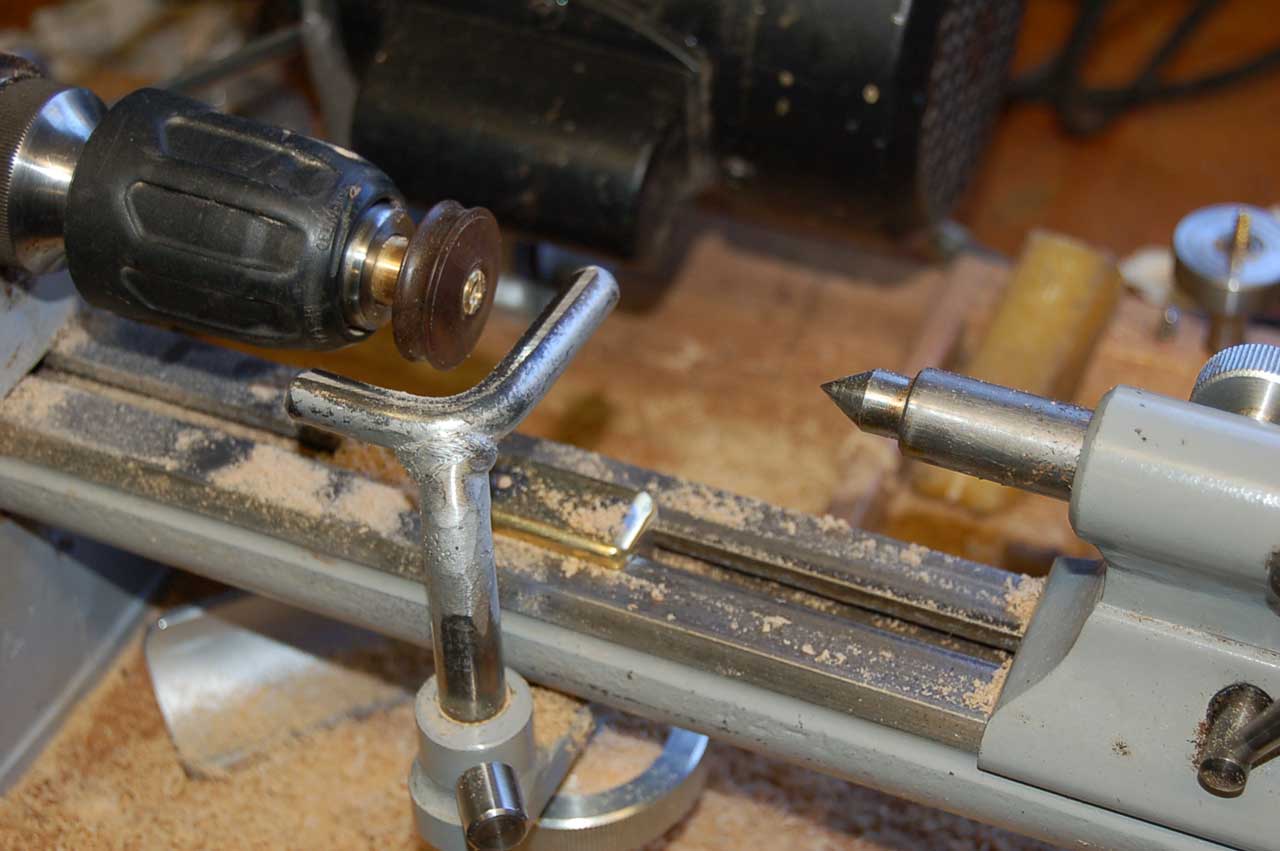
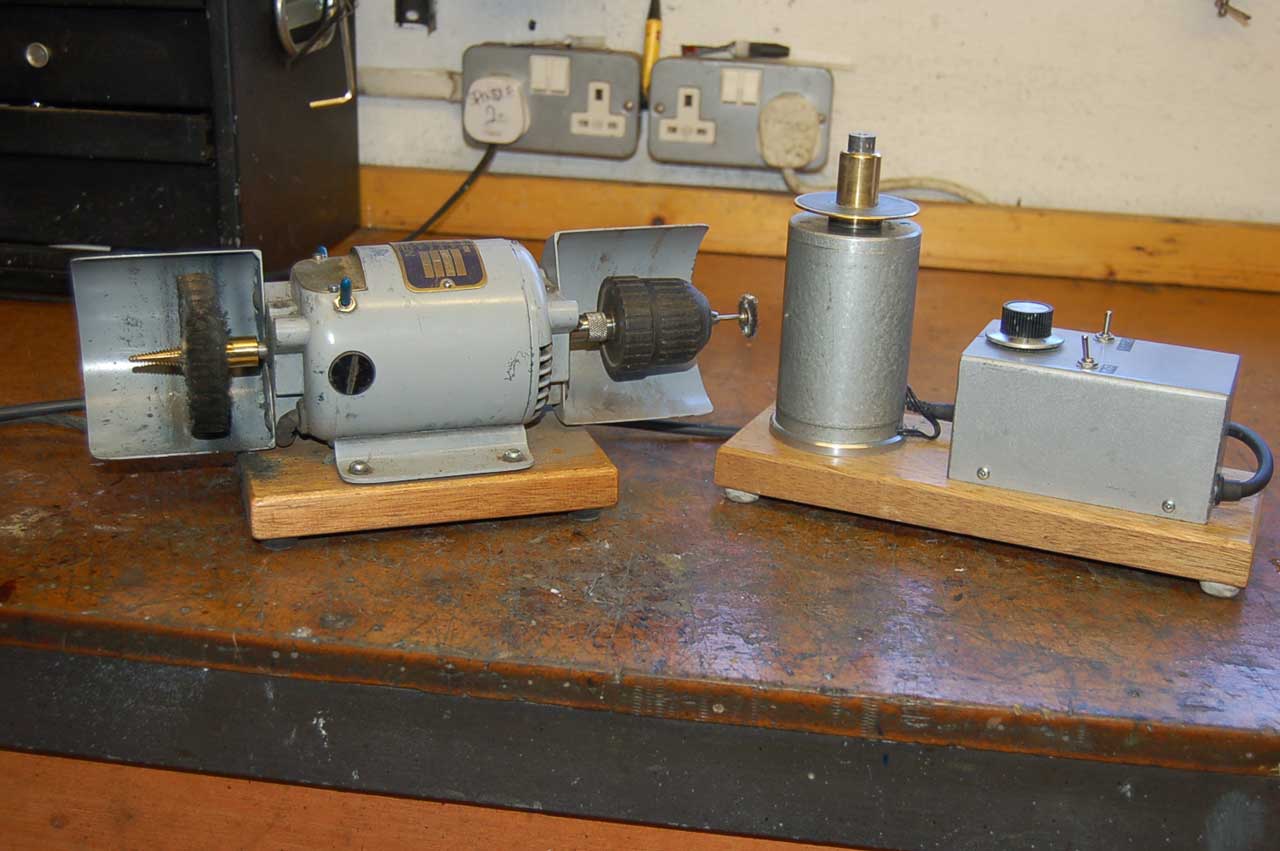
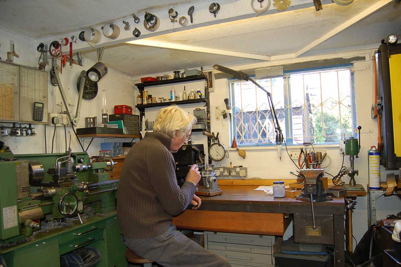
Photos of Christopher Kirby's Shop
Overall
Swaging Press
Wood Lathe
Stock & Custom Lens polishing
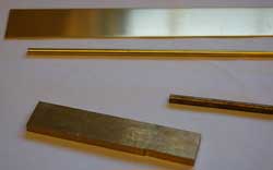
Brass & Copper Stock
Cutting glass cylinder from window pane
Lens Grinder
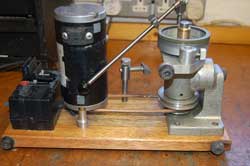
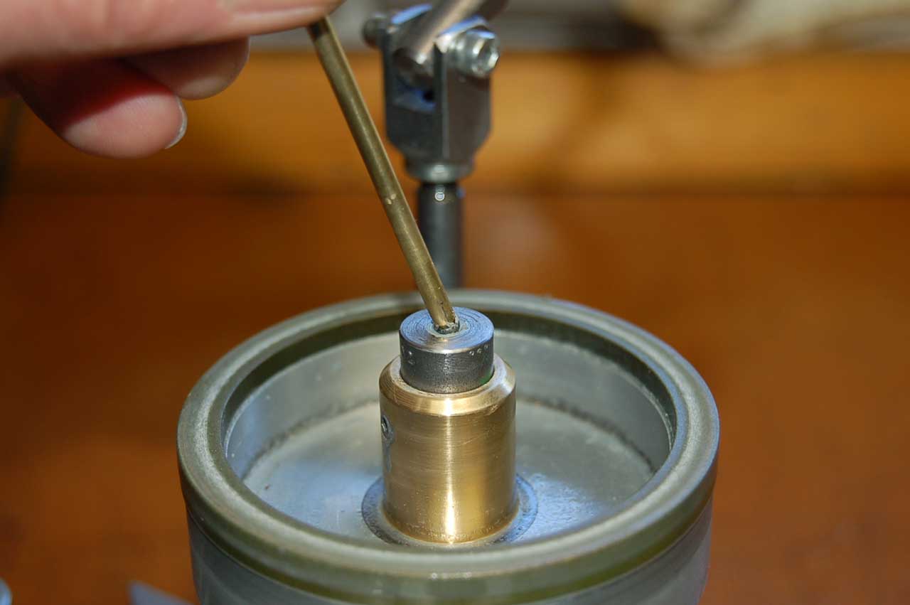
Fig 5 Grinding Lens
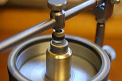
Fig 6 Concave tool shapes lens
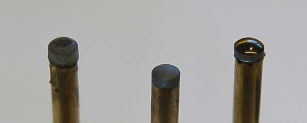
Grinding a Lens
Assortment of lenses of different sizes
Different stages of grinding - polishing
YouTube,Objectivity, Lost Microscopes -
The Delft School of Microbiology - A. van Leeuwenhoek -
Making a Van Leeuwenhoek Microscope Lens, Making an Antoni van Leeuwenhoek microscope replica by Hans Loncke, the Netherlands -
Museum Boerhaave - Van Leeuwenhoek replica -
YouTube: The Gadget That Changed How We See The World | Cell | BBC Earth Science, 4:35 -
This is a lens that looks like a glass rod with a spherical end and a flat end. The spherical lens comes to focus at the flat end. If a small photograph is mounted on the flat end and you look into the spherical end you will see a magnified image of the photograph. This would be a device that could be used by a spy to read a microdot (Wiki), or a small message on film like in the Spy Nickel.
According to Wiki these are a type of Stanhope (Wiki) lens.
University of Arizona, College of Optical Sciences, Stanhope Magnifier - Flat end rests on book and spherical end towards your eye.
Class 359 Optical: Systems and Elements /
362 Compound Lens System /
368 Microscope
359/644 Spherical Lens (no microscopes found)
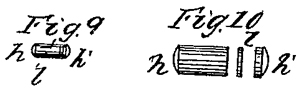
33031 Portable Microscope, Rene P. P. Dagron France, Aug 13, 1861, 359/368; 63/23 - Bullet lens - oldest patent in US class 359/368 i.e. oldest U.S. microscope patent.
Fig 9 & 10 Eyepiece (h) focuses on object (l). Object glass (h') the purpose of which I'm not sure (maybe condenser for light source).
203323 Improvement in finger-rings, William B. Closson, May 7, 1878, 63/1.12; 40/639; 63/15 - need to remove ring to get light from finger side
752889 Portable Microscope and case therefor, Feb 23, 1904 - looks like compound scope class 359/368
2492691Illuminated world globe, William H Dietz, Dec 27, 1949, 434/145, 362/809 - With Stanhope images of cities, &Etc.
4416074(toy plastic filmstrip) Ring viewer, Benjamin G. Guerrero, Saint Elmo B. Berford, Mattel, Inc., Nov 22, 1983, 40/364, 63/1.12, 63/23, 40/661 - good list of references
YouTube: Stanhope Viewers: Victorian Portable Peepshows, 10:49 - @6:41 use in war & espionage
Microdots
The Stanhope magnifier was used in the Civil war to view small photos where the magnifier and lens were concealed inside a Minié ball (Wiki).
See page 10 of Ref 2.
J. Edgar Hoover (Wiki) wrote a report on W.W.II German spying where he says "...Professor Zapp, inventor of the microdot process, at the Technical High School in Dresden." Which was wrong, the microdot was invented by Emanual Goldberg (Wiki). Hoover may have been thinking about Walter Zapp the inventor of the Minox camera.
Minox (Wiki) patents
Minox: the OG Spy Camera, 23:00 -
2147567 Lens mechanism for photographic apparatus, Walter Zapp, Dec 24, 1936, Valsts Elektrotechnika Fabrika, 396/144, 74/459.5, 359/823, 74/424.5, 74/89.45, 74/417 - Minox
2169548 Roll film camera, Walter Zapp, Dec 22, 1936, Valsts Elektrotechnika Fabrika, 396/401, 396/472, 396/535, 396/448, D16/212 - Minox (Wiki)
3409343 Magnifying viewing device, Zapp Walter, Oct 23, 1965, 359/431, D16/135 - Not clear where this is used.
McMaster-Carr - 8996K21 Heat-Resistant Borosilicate Glass Balls,
3/32" Diameter, packs of 50
3/32" (2.4mm) dia Winsted 3200940F1ZZ00A0
McMaster-Carr 8996K21
This might be the condenser lens for the FoldScope?
In the Stanford Foldscope POLS (Ref 1) article there's a 3D graph showing ball diameters between 100 and 1200 um -vs- Index of Refraction between 1.9 and 1.4 resulting in resolutions in the range of 0.6 to 2.2 um.
The FoldScope lenses are 140X and 430X with resolutions of 2 um and 1.4 um respectively.
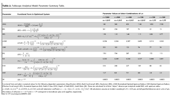
Table 2 from
POLS - Foldscope: Origami-Based Paper Microscope - Full document.pdfLists optical characteristics of a number of commercially available ball lenses.
Note elsewhere in the article they claim up to 2180X without immersion oil. In Table 2 (see below) the NA varies between 0.2 to less than 0.5 hence no need for oil.
Ball Lenses (reformatted for this web page from POLS article)
The ball lenses used in constructing Foldscopes included material types borosilicate, BK7 borosilicate, sapphire, ruby, and S-LAH79. The vendors included:
Swiss Jewel Co: 300 µm sapphire lens (Model B0.30S) & 300 µm BK7 borosilicate lens Model BK7-0.30S & 1.0 mm BK7 borosilicate lens Model BK7-1.00
Edmund Optics:
Winsted Precision Ball (McMaster-Carr): 2.4 mm borosilicate lenses p/n: 3200940F1ZZ00A0 (I have these, see above)
Note that half-ball lenses from both Edmund Optics and Swiss Jewel Co. were also tested for use as condenser lenses for the LEDs
Summary of Table 2
1200
500
400
150
150
100
n
1.517
1.517
1.517
1.517
1.77
1.77
borosilicate glass
sapphire MAG
140
340
430
1140
1450
2180
RES (um)
1.90
1.52
1.44
1.13
0.86
0.77
NA1
0.200
0.249
0.264
0.337
0.444
0.491
Max MAG2
200
249
264
337
444
491
Click on table below for larger .pdf version.
Note 1: NA as reported in the PLOS paper
Note 2: based on 1000 * NA - Notice that all except the lowest power glass ball have "hollow magnification". That's to say they are actually not resolving the details.
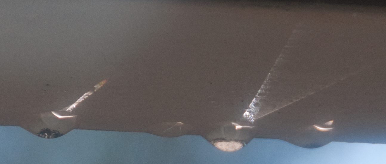
Morning of 3 Nov 2016 - Sun back lighting the forest and water drops on the bottom of the rain gutter.
In the drop at the left in the close up (click image below for close up) the bottom of the drop appears dark, but a closer look will show that you're seeing the tops of trees.
The drop to the right sees the sky and is light colored.
Microscopes
Labophot Microscope, Nikon SMZ-U Stereo
Mitutoyo Toolmakers Measuring Microscope 176-134
Multiphot System, Nikon microscope
Omnicon 3800 Tumor Colony Analyzer (TCA) Automated Inverted Biological Microscope
Unitron Auto-Illumination Inverted Microscope Mica-U3X MiC3-244
Unitron No. 83444 Microscope
Digital Photography 202: Close Up, Macro & Micro
Digital Photography 206 Micro Photography - includes using microscope objectives on camera (sort of simple photo microscope)
Optical Spectrum Analyzers
Monolight- rotating optical gratingLight
Beseler PM1 Darkroom Color Analyzer - variable color filters in front of photomultiplier tube (Wiki PMT)
Wollensak L3524D Direct Vision Spectroscope - wavelength scale can be calibrated using table salt in a flame.
Ocean Optics HR2000 Spectrometer - coverts 467 nm to 670 nm i.e. uses the H9 grating and a linear CCD sensor, USB interface to computer
Optics
Electro-Optical Gadgets
LED
M18 IR Binocular
PAS6 Metascope IR Viewer & IR Source
IR Beacon
M227 Signal Lamp Equipment SE-11
UAS-4 Infrared Surveillance System, AN/AAS-14 Infrared Detecting Set, MK-898/AAS-14A IR Optical Filter Kit
Theodolites -
Leitz 115 Transit -
Del Mar Photonics may make the Foldscope lenses, but so far I haven't been able to buy any from them. Still trying. Greyhawk Optics is related to Del Mar Photonics.
Stanhope MicroWorks, Stanhope Jewelry, Pen Peep - all related.
Stanhope Info - also called peeps. has book
Antique Microscopes - Microscope-Related United States Patents: 1853-1915 -
Aperture-Embedded Polymer Microlenses for Ultra-Low-Cost Microscopy Platforms (Foldscope) By Laurel Kroo , George K. Herring & Manu Prakash all from Stanford
Ref 1 POLS - Foldscope: Origami-Based Paper Microscope - Full document.pdf
McMaster-Carr - 8996K21 Heat-Resistant Borosilicate Glass Balls, 3/32" Diameter, packs of 50
Instructables - Cardboard van Leeuwenhoek microscope
To Make a Van Leeuwenhoek Microscope Replica 5/16/96 copyright Alan Shinn - parts drawings
Fun Science Gallery - A Glass-Sphere Microscope by Giorgio Carboni - power of ball lens = 340/d where d=diameter in mm.
Ref 2 the Microdot history and application by William White 1992, foreword by Ray Cline - appendix A Peersonal Biographical Memoir by Walter Zapp 1981 about the development of the Minox camera
Ref 3. Neutron tomography of Van Leeuwenhoek’s microscopes,